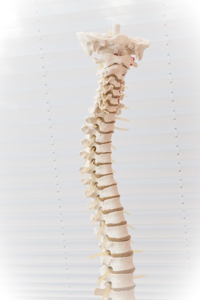
Lumbar and cervical disc herniation. 80% of people say that they suffer or have suffered from prolonged back problems. In older people, most of the time they are produced by arthrosis, by a contracture or by a bad posture and not by a hernia. But, sometimes the pain is caused by this one.
In order not to confuse it with other ailments, the Nomenial team answer the most frequent questions about it:
What is Lumbar and cervical disc herniation?
Herniated disc is a type of injury that affects the discs of the spine.
The spine is formed by several parts, among which are the invertebral discs. These are the tissues between the bones of the spine.
Composed of a soft center with a gel-like texture and a more solid outer covering, they form a joint between each of the bones. This joint cushions the movements that occur between two adjacent vertebrae and allows the bones to move.
As we age, the spinal discs deteriorate and lose flexibility and elasticity. When the outer covering of the disc tears, the soft center can move out of its normal boundaries. The release of this material is known as a herniated disc.
Risk factors for Lumbar and cervical disc herniation
Sex: Men between the ages of 30 and 50 are more likely to have a herniated disc.
Sedentary life: Performing gentle daily exercise decreases the occurrence of inflammatory processes in the discs.
Obesity: Being overweight increases the pressure on the discs in the lumbar area.
Carrying weight improperly: Lifting heavy objects using the back instead of the strength of the legs can cause a herniated disc. If you turn your back at the same time as you are lifting, the damage may be greater.
Repeatedly performing activities that overload the spine: Work that requires physical effort that involves stretching or bending causes the spine to deteriorate. To avoid this, safe movement and loading techniques can be used.
Frequent driving: Sitting for long periods of time, coupled with the vibration of the vehicle engine, can put increased pressure on the spine and discs.
Smoking: Smoking is believed to decrease the oxygen supply to the disc and cause faster disc degeneration.
Genetics: There are hereditary factors that make the early appearance of a herniated disc more likely.
Types of disc herniation: lumbar and cervical
Lumbar disc herniation
They are the most frequent. The hernia occurs as a result of the output of the contents of the interior of a disk in the lower spine, which causes low back pain, also known as low back pain.
Herniated cervical disc
The affected discs are those in the neck area (cervical spine).
Symptoms of Herniated Disks
The main and general symptom is intense pain in the area where the hernia is located.
Depending on the type of hernia, the other symptoms are as follows:
Sciatica due to disc herniation: Pain and/or numbness in one or both lower extremities (radiates from the lumbar and is directed from the buttock downwards).
Difficulty in performing everyday movements such as moving, bending or getting up and turning over in bed.
You may experience a feeling of weakness or numbness in your buttocks and/or lower extremities (both in the back or front and inside one of your legs).
Symptoms of Herniated Cervical Disc
Pain in the back of the neck or neck.
Difficulty in performing actions such as moving the neck or lifting the arms.
You may experience a feeling of weakness or numbness in one of your upper extremities.
In extremely rare cases where a herniated disc with severe symptoms is experienced, the so-called ponytail syndrome can occur: loss of bladder or bowel control, resulting in an inability to hold urine or stool.
But, as we pointed out, it only happens in cases of large fractures or massive hernias, so it is not a common symptom.
Characteristics of disc herniation pain
The pain is felt right in the region of the back where the hernia has occurred. That is to say, if the hernia is cervical, a deep pain will be felt in the back of the neck.
The sensation of pain usually appears immediately or several hours after the effort or trauma.
This affliction usually becomes more acute when making certain movements or efforts and when doing actions such as coughing.
This initial discomfort may improve after a few days, but it usually worsens later, causing increasingly frequent, longer and more painful episodes.
Herniated disc stages:
Disc degeneration: Corresponds to the first stage. The nucleus pulposus is weakened but in a very slight way, so it does not come to produce the hernia.
Prolapse: The disc changes its shape or position. It begins to form a small lump or bulge that may surround the spinal cord.
Extrusion: The gelatinous nucleus crosses the external cover but without coming out and losing contact with the disc.
Sequestration: It occurs during the last stage. The nucleus pulposus even comes out of the disc.
Diagnosis of hiscal hernia
The diagnosis of a herniated disc is based on a review of the patient’s medical history.
After that, the first examination that the doctor will perform will be a physical examination of the spine and the upper and lower extremities. Through this he will check the flexibility, the range of movement and all those signs that warn him of the existence of a herniated disc.
Along with the physical examination, he will also perform a neurological exam to detect if there is muscle weakness. To check it, the doctor will require the patient to walk supported by the heels and toes.
In addition, he will analyze the muscular reflexes. These, as a result of the damage caused by the hernia may have been reduced or, in more serious cases, even disappeared. To do this, it will test if the patient, by executing a soft touch on the knee and ankle, reacts to this action.
The specialist may then also ask the patient to perform certain daily movements to check whether he or she is performing them with difficulty, and to observe how the spine responds to them.
For example: sitting, walking, moving the neck, shoulders or hands, or bending over to each of the four sides. Finally, to confirm the existence of a herniated disk, the physician may prescribe a magnetic resonance imaging (MRI) study.
This study may be one of the following:
Electromyography: It will show which is the affected nervous root and where it is compressed.
Myelography: It will limit the size and location of the herniated disk.
MRI: It will determine if the hernia is putting pressure on the spine.
Spinal X-ray: It will serve to rule out other conditions that present symptoms very similar to those of a herniated disk.
Treatment of herniated disk:
Non-surgical treatment:
Medications
Analgesics: If the pain is not acute, but remains between mild and moderate, the specialist may recommend taking analgesics. Along with these, he may prescribe non-steroidal anti-inflammatory drugs.
Cortisone injections: If the pain is not relieved by oral medication, the doctor may prescribe a corticosteroid to be injected into the area around the spinal nerves. Imaging tests may indicate the exact location of the injection.
Muscle relaxants: If the patient suffers muscle spasms, they may prescribe relaxants that will help relieve the pain.
Therapy
Physical therapy can reduce pain and improve a patient’s strength and flexibility. Physical therapists will design a plan of exercises and positions with which the patient will strengthen the muscles of the back, abdomen and legs.
Along with these anaerobic exercises, other aerobic exercises such as walking or exercising on a stationary bike may also be prescribed.
With all of this, the patient will not only reduce his or her pain, but will also gain autonomy and mobility.
Surgical treatment:
If the discomfort does not improve with conservative treatment, after a reasonable time the possibility of surgical treatment will be assessed.
However, this is not the most common solution, as only 10% of herniated disc patients end up needing surgery.
The surgery practiced is minimally invasive and consists simply of the extraction of the hernia.
How long does a disc herniation operation last?
As for the time the patient is in the operating room, the operation lasts between 1
and 2h and can even be performed under epidural anesthesia.
As far as recovery is concerned, it usually takes between 2 and 6 weeks.
